
AI AFib Detection Accuracy: Evidence and Clinical Impact
Rhythm360 breaks down AI AFib detection accuracy, false positives, and clinical evidence. See how to turn AI insights into reliable patient care.

Key Takeaways
- Pooled AI AFib detection accuracy reaches 90–97% sensitivity and 95–99% specificity in controlled studies, yet these figures describe study-level performance rather than per-patient reliability in monitored populations.
- Paroxysmal AFib episodes are frequently missed by single-snapshot ECGs, and advanced sinus-rhythm prediction models still show false-negative rates of approximately one-third in prospective validation.
- False-positive alerts from artifact, ectopy, and single-lead limitations create substantial clinical workload and patient anxiety, especially in low-prevalence screening settings.
- AI performance matches or exceeds junior clinicians on retrospective datasets but does not consistently surpass experienced cardiologists who integrate broader clinical context.
- Rhythm360 unifies CIED and RPM data, applies AI-powered triage, automates documentation and billing, and turns AFib detection into actionable, billable clinical care.
Schedule a Demo with Rhythm360
The Problem: Pooled Accuracy And Real-World Reliability
The table below summarizes frequently cited study-level evidence on AI AFib detection accuracy. Each figure comes from a specific population, validation protocol, and signal modality, and each carries a limitation that pooled headlines usually omit.
Study / SourcePopulationSensitivity / SpecificityStated LimitationManetas-Stavrakakis et al., J Clin Med, 2023 (meta-analysis)Multi-setting; single-lead AI-ECG92.3% sensitivity / 96.2% specificity (pooled); outpatient: 90.7% / 98.1%Pooled figures span heterogeneous datasets; accuracy maintained with accessible AI devices but not validated across all monitored populationsAI-enabled PPG systematic review and meta-analysis (Frontiers in Cardiovascular Medicine, 2026)Smartwatch / wearable PPG; general and clinical populations~95.1% sensitivity / 96.2% specificity (pooled PPG); BASEL device-level PPG: Apple 89%, Samsung 85%, Fitbit 82%, Withings 79%Real-world accuracy lower due to motion artifact, ectopy, and signal degradation; PPG cannot capture P-wave morphologyMayo Clinic sinus-rhythm prediction model (Attia et al., Lancet, 2019; reviewed in Pantelidis et al., Biomedicines, 2026)649,931 sinus-rhythm ECGs; 180,922 patients; internal test setAUC 0.87 (single ECG) / ~0.90 (aggregated ECGs); sensitivity ~79% / specificity ~79%Retrospective, single-system; ground truth includes AF within 31 days of ECG, blurring latent detection and forecasting; most AI-AF studies remain retrospective
Pooled accuracy figures describe study-level performance, not per-patient reliability. A 2026 systematic review in Discover Artificial Intelligence (Volume 6, article 977) analyzed 75 studies on machine learning and deep learning for ECG-based cardiovascular disease prediction and found that diagnostic performance varied substantially by dataset characteristics, preprocessing strategy, model architecture, validation protocol, and evaluation metrics. Pooled figures therefore cannot transfer directly across study populations or clinical settings. The same review reported that many studies showed excellent internal validation performance, yet relatively few performed external or prospective clinical validation, which the authors identify as a major barrier to routine clinical adoption.
With those study-level caveats in mind, clinicians still need a clear view of the broader accuracy figures that shape expectations.
How Accurate Is AI at Detecting AFib?
Across published meta-analyses, AI-ECG systems detect atrial fibrillation with pooled sensitivity generally between 90% and 97% and pooled specificity between 95% and 99%. The largest 2026 meta-analysis reported a pooled sensitivity of 92.6% and specificity of 99.1%, depending on lead configuration, signal modality, and study population. Wearable PPG-based systems report similar pooled ranges, and the Mayo Clinic sinus-rhythm prediction model achieves an AUC of 0.87 to 0.90.
These figures come from controlled validation datasets that are often retrospective, single-system, and drawn from benchmark ECG libraries such as MIT-BIH, PTB-XL, and PhysioNet. The 2026 Discover Artificial Intelligence systematic review confirms that the most frequently used datasets in AI-ECG research are MIT-BIH, PTB-XL, PhysioNet, CPSC, and UK Biobank. These populations differ from the heterogeneous, comorbid patients in a typical monitored cardiology panel.
How Accurate Is AI at Detecting Paroxysmal AFib?
While pooled figures describe overall performance, paroxysmal AFib presents a distinct challenge that single-snapshot detection cannot overcome. Paroxysmal AFib defeats single-snapshot detection by design. An episode that lasts minutes or hours, often during sleep, will not appear on a 10-second ECG taken during sinus rhythm. Continuous monitoring addresses this limitation, which pooled accuracy figures do not capture.
The Mayo Clinic model changes the math by predicting paroxysmal AF from a normal sinus-rhythm ECG. The Attia et al. convolutional neural network, trained on 649,931 sinus-rhythm ECGs, achieved an AUC of 0.87 with a single ECG and approximately 0.90 when aggregating multiple ECGs per patient. The model identifies hidden atrial substrate before an episode is ever recorded. However, a 2026 narrative review by Pantelidis et al. in Biomedicines (published May 7, 2026) concluded that most AI-AF studies remain retrospective, single-system, and focused on diagnostic yield rather than clinical outcomes such as stroke or mortality. The authors call for robust prospective trials before routine integration of AI-guided AF prediction into clinical decision-making.
The paroxysmal AFib detection literature also highlights a practical false-negative problem. A 2026 single-center prospective observational study at Oita University Hospital (Sato et al., Journal of Arrhythmia, doi:10.1002/joa3.70439) enrolled 104 adults with documented paroxysmal AF admitted for catheter ablation and found that approximately one-third of patients, 36 of 104, were classified by AI-ECG into the lower AF likelihood group during sinus rhythm. Sato et al. concluded that AI-ECG-based AF likelihood assignment is heterogeneous among patients with documented paroxysmal AF during sinus rhythm and that patient characteristics such as hypertension and left ventricular mass index should be considered when interpreting AI-ECG output. Continuous monitoring substantially increases capture probability compared with episodic snapshots. False negatives can still occur, however, in patients whose atrial substrate does not appear in the features a given algorithm was trained to detect.
For practices building a protocol around AFib CIED detection, the implication is direct. Clinicians should interpret algorithm output in clinical context rather than treat it as a binary screen.
What Causes False Positives in AI AFib Detection?
False negatives represent only half of the accuracy challenge. False positives create their own clinical and operational burdens, and understanding their causes helps teams manage alert volume.
The following are the primary technical causes of false-positive alerts in AI-based AFib detection systems:
- Artifact and baseline wander, which disrupt RR-interval regularity and mimic AF in automated analysis
- Ectopic beats, including premature atrial contractions (PACs) and premature ventricular contractions (PVCs), which produce irregular pulse intervals that basic PPG algorithms cannot distinguish from AF
- Sinus arrhythmia, particularly in younger patients or athletes, where physiologic RR variability triggers irregular rhythm notifications
- Single-lead limitations and the absence of P-wave morphology in PPG-based systems, which prevents differentiation of true AF from mimicking arrhythmias
- Degraded signal fidelity in individuals with darker skin pigmentation or cutaneous conditions such as tattoos and scars, which can reduce PPG accuracy
Translating specificity into operational reality requires simple arithmetic that pooled figures obscure. Even a specificity of 99% applied to a monitored panel of 1,000 patients generates approximately 10 false-positive alerts among patients without AF. Each alert typically requires confirmatory workup, including extended ambulatory monitoring, cardiology consultation, and anticoagulation risk discussion, which consumes clinical time and generates patient anxiety. Chen et al. document that false-positive alerts from consumer-grade AF detection devices provoke patient anxiety and trigger cascades of unnecessary evaluations, including specialist consultations and supplementary testing, imposing a considerable financial burden on healthcare systems. An analysis in the European Journal of Preventive Cardiology confirms that positive predictive value falls as baseline prevalence falls. The false-positive burden therefore peaks in the very settings where screening feels most attractive: low-prevalence, younger, or lower-risk populations. For a deeper look at managing alert volume, see AI Triage Heart Failure Alerts: Cut Alert Fatigue by 80%.
AI And Human Clinicians: How Accuracy Compares
AI-ECG systems match or exceed junior clinician performance on retrospective datasets, particularly for pattern recognition in clean, well-labeled ECG libraries. The advantage narrows or reverses when experienced cardiologists serve as the comparator. A 2026 narrative review in the International Journal of Emergency Medicine by Khandelwal et al., synthesizing 122 studies published between 2015 and December 2025, found that AI-ECG systems achieve diagnostic performance comparable to or exceeding junior clinicians on retrospective datasets but do not uniformly outperform experienced cardiologists or emergency physicians. The ROMIAE prospective multicenter study illustrates this gap. The DAMI-ECG model achieved an AUROC of 0.915 during retrospective external validation, which declined to 0.814 in prospective deployment across multiple emergency departments.
The explanation is structural. AI-ECG evaluates electrical waveforms. Experienced clinicians integrate ECG findings with comorbidities, medication history, prior tracings, and bedside assessment, context that no current algorithm ingests. AI therefore performs best as decision augmentation rather than autonomous replacement of clinical judgment.
Device Variation, Smartwatches, And Usability
In controlled studies, wearable ECG sensitivity for atrial fibrillation detection varies by device and study. A systematic review of ECG patches reported 92–98% sensitivity, while individual device studies ranged from about 82% to 100%. Real-world performance diverges further. The 2026 BASEL wearable study of 201 patients with atrial high-rate episodes or known AF found PPG sensitivity of 89% for Apple Watch, 85% for Samsung, 82% for Fitbit, and 79% for Withings, while single-lead ECG diagnostic accuracy exceeded 95%. Pooled numbers hide these differences entirely.
Apple Watch AFib Accuracy The Apple Heart Study, a prospective single-arm study of 419,297 participants, found that 0.52% received an irregular pulse notification, with a positive predictive value of 84% for AF on a concurrent ECG patch. AF was confirmed in only 34% of notified participants who completed follow-up. That figure reflects both the PPV limitation and the challenge of follow-up completion in a general-population screening context.
Why ChatGPT Cannot Read ECGs Reliably Large language models such as ChatGPT currently have no validated diagnostic role in ECG interpretation. They are not cleared by the FDA for rhythm analysis, have not been prospectively validated against clinical outcomes, and should not replace validated AI-ECG algorithms or clinician review.
A 2026 randomized comparison in Europace found that only 45.3% of participants met the predefined threshold of at least 24 hours of analyzable ECG across self-applied patch systems, with the main barrier being device application and activation before signal acquisition rather than rhythm-detection accuracy. Device usability therefore becomes the primary determinant of monitoring yield. For a decision framework on selecting among AFib remote monitoring devices, operational factors matter as much as published sensitivity figures.
These operational realities, including fragmented data, alert burden, and workflow gaps, define the environment that any unified monitoring platform must address.
The Solution: Operationalizing AI AFib Detection In A Monitored Population
Detection marks the beginning of the workflow rather than the end. An AI-generated AFib alert gains clinical value only after teams triage, review, document, and, when appropriate, bill for the associated care. The gap between pooled algorithm accuracy and real-world detection yield therefore reflects operational constraints as much as technical performance.
Rhythm360 is a vendor-neutral, HIPAA-compliant, AI-powered platform that closes this gap. The platform unifies CIED and RPM data from all major device manufacturers into a single source of truth. This structure eliminates fragmented OEM portal workflows that create data silos and administrative overload. AI-powered alert triage filters non-actionable noise and prioritizes clinically significant events, so a new-onset AFib alert on a Saturday morning reaches the right clinician in time to initiate anticoagulation before a stroke occurs.
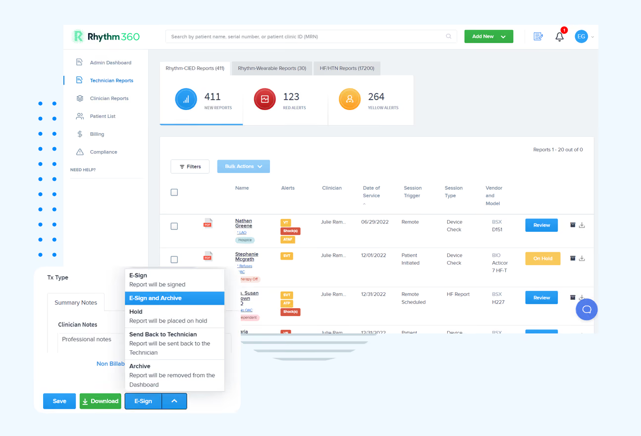
Rhythm360 automates reporting and billing documentation for the full spectrum of remote monitoring CPT codes. The platform supports the full range of remote monitoring CPT codes, but the billing rules differ by device type. Pacemakers and ICDs follow a 90-day cycle: 93294 and 93295 cover the professional components, while 93296 covers the technical component. Physiologic monitors such as CardioMEMS and subcutaneous cardiac rhythm monitors follow a 30-day cycle instead, using 93297 and 93298 respectively. Both 30-day codes can be split into professional (-26) and technical (-TC) components, which matters for practices that bill separately for each. Automated CPT tracking prevents device-type mismatch denials and unbilled technical components, helping practices recover previously lost revenue.
Practices implementing Rhythm360 have achieved up to an 80% reduction in critical alert response times and up to a 300% increase in revenue capture and profitability. For a broader view of how AI-powered platforms are reshaping cardiac monitoring workflows, see AI In Cardiac Telemetry: Reduce False Alarms And Unify Data and the 2026 Buyer's Guide To AI-Powered Cardiac Implant Monitoring.
Evidence Gaps And Open Questions
The evidence base for AI AFib detection accuracy still contains meaningful gaps that pooled figures cannot resolve. Generalizability across diverse populations, including patients with hypertension-related atrial remodeling, darker skin pigmentation, and multiple comorbidities, remains incompletely established. Performance in low-prevalence screening settings is systematically more modest than headline figures suggest because positive predictive value falls with prevalence regardless of specificity. Standardized real-world benchmarks for AI-ECG performance do not yet exist. The 2026 Discover Artificial Intelligence systematic review found that relatively few of the 75 included studies performed external or prospective clinical validation. The authors identify this gap as the primary barrier to translating AI-assisted ECG models into routine clinical practice.
Practices building protocols around AI AFib detection should treat published accuracy figures as a starting point for evaluation within their own patient population. For more on how AI filters noise in practice, see How AI Filters Noise in Remote Cardiac Monitoring.
Conclusion: Turning Accuracy Into Reliable Care
Pooled AI AFib detection accuracy is high, but it cannot serve as the sole basis for a clinical protocol. The gap between a 92%–95% pooled sensitivity figure and the detection yield in a real monitored population, shaped by paroxysmal burden, comorbidities, signal quality, and algorithm architecture, defines the practical risk that practices must manage. An effective solution framework unifies CIED and RPM data, applies intelligent triage to separate actionable alerts from noise, generates auditable documentation, and supports compliant billing across the full remote monitoring CPT code set. Rhythm360 is built to close that gap.
Start Closing Your Detection Gap
.avif)